
Imaging Core
PI: Andy Alexander
Co-PI: Rasmus Birn
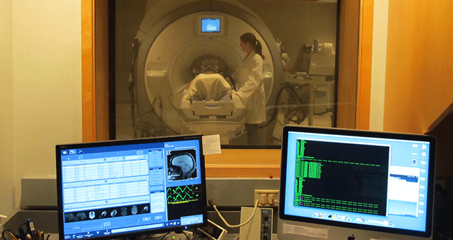
The Imaging Core provides support for the acquisition, analysis and interpretation of magnetic resonance imaging data that is being collected in the Center Projects. More specifically, the Imaging Core is developing and maintaining pulse sequences and protocols used in the Projects, and is also responsible for ensuring high and consistent image quality. Imaging protocols for human infants and adolescents, and very young nonhuman primates are employed for structural, microstructural (diffusion tensor imaging), functional (both resting/sleeping and task-based) mapping of the brain. The core is developing and maintaining image analysis pipelines used in the projects. These include methods for analyzing structural morphometry (including amygdala and prefrontal volumes), diffusion tensor imaging measures of amygdala, limbic and prefrontal white matter pathways, task-based functional MRI measures and resting-state functional connectivity measures. Selective strategies for analysis in very young monkeys and human infants are being developed to facilitate measurements in the young brain with immature myelination. Finally, the core is helping to evaluate and implement new image acquisition techniques and image analysis strategies that the Center investigators deem to provide novel useful and/or more accurate measurements.
Contact our Webmaster with any Questions or Feedback
© 2014 The Board of Regents of the University of Wisconsin System


